
The world of the very vast has been opened up via optical telescopes which focus ancient photons through glass lenses, radio telescopes which receive non-visible electromagnetic signals from distant stellar bodies, and a variety of gamma-ray, microwave, and infrared telescopes, all of which allow us humans to "see" far into the deepest reaches of space, even as far as 13-14 BILLION years ago. Imagine the wonder felt by Galileo Galilei when he fixed his crude but effective telescope device upon Saturn, and saw something similar to this. It probably blew his mind, and it was so heretical to most that many refused to look through his telescope altogether, for fear of offending their creator.

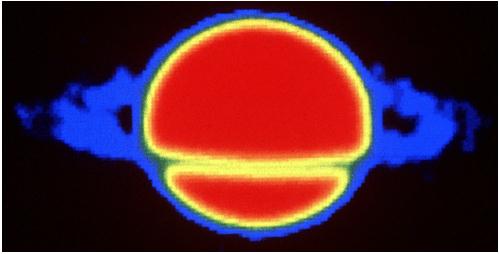
Here is a radio telescope image of the same planet. Through different energy wavelengths, different features and properties of stellar objects come into focus.
The world of the very small was discovered through optical microscopes, which allowed humans to see for the very first time, the unbelievably vast amount of teeny tiny life-forms that we share our world with, from tiny crustaceans to single-celled organisms to bacterial life. These creatures are everywhere, and go about their life-processes with most of humanity completely oblivious to them. Here is a microscopic image of a single-celled paramecium. Note that even though this is a "simple" organism, the structures that make up it's "body" are just as complex as anything we have in our large "complex" bodies. Can you imagine the awe that was felt by the very first humans to lay eyes upon such things?

After a few centuries, and once technology had developed to allow it, humans created a device which allowed for the resolution of tiny details on these creatures, as well as the viewing of structures and viruses which are hundreds of times smaller than the smallest bacteria, the scanning electron microscope. This device actually rained a beam of electrons, mass-less or nearly mass-less negatively charged particles, onto the surface of what was being viewed, resulting in incredibly detailed three-dimensional black and white images. Below is an image of lactic acid bacteria, on the scale of nanometers (millionths of a meter) created by scanning them with the beam of electrons.

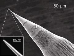
There have been even greater advances in microscopy that the scanning electron microscope. In 1981 the Scanning Tunneling Microscope was developed by scientists at IBM Zurich. It is a very complicated device that works, not on light or beams of electrons, but on what is a very strange feature of quantum mechanics. By using a nearly infinitesimally tiny needle tip instead of a beam of electrons, and measuring the "interference" between the tip's electron cloud and the electron cloud of the item being magnified, actual images of atoms and molecules are possible. On the quantum level these fields interact as if "tunneling" into the surface of what is being studied, for in the quantum world, energy and matter are but two sides of the same coin. These "needles" are so sharp that the ends of them are one to two atoms thick! Insane!!!! Here is an image of an etched STM tip, in nanometer scale.
The images produced by the STM seem almost mystical, for they show the raised electron clouds of the individual atoms. The STM can even be used to move individual atoms around, as in this image. Each raised bump is an individual atom. The "ripples" seen are the interference patterns between the atoms.

Scientists have developed a new imaging technology that produces the best three-dimensional resolution ever seen with an optical microscope. With this new tool, scientists can pinpoint fluorescent labels in their images to within 10-20 nanometers – about ten times the size of an average protein – in all three dimensions. The researchers say they now have an extremely powerful technology that will help reveal how biomolecules organize themselves into the structures and signaling complexes that drive cellular functions. ( Science Daily )

The above image is an example of what the new microscopy can show. It is interferometry combined with the super-high resolution photoactivated localization microscopy, or iPALM for short.
The three-dimensional distribution of membrane proteins within a cell revealed through iPALM imaging. The vertical position of fluorescently labeled VSVG proteins has been color coded, with red molecules being the deepest and purple the highest.We humans are some bad-ass mo-fucks. -FUPPETS- loves when science kicks out the jams.
No comments:
Post a Comment